Treatment
Bone Graft for Dental Implants — What It Is and When You Need It
A bone graft is a surgical procedure that adds bone material to the jaw to create sufficient volume for a dental implant (the titanium fixture placed in the jawbone) to be placed and integrate securely.
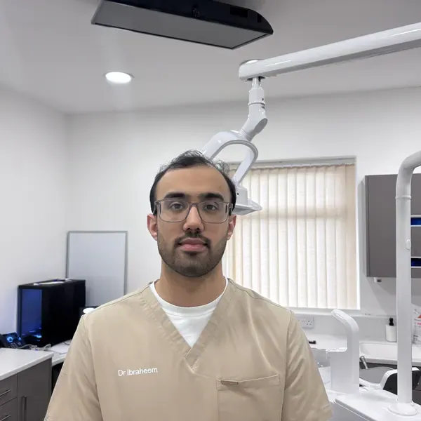 Clinically reviewed by Dr Ibraheem Ijaz, Principal Dentist · GDC 301711 · 500+ implants placed · Last reviewed August 2026
Medical disclaimer: This page is for general information only. It does not replace an examination or treatment by a dentist.
What is a bone graft for dental implants?
For an implant to function reliably, it must be surrounded by adequate bone on all sides. When a tooth is lost or extracted, the jawbone beneath it begins to undergo alveolar ridge resorption — a natural process in which bone volume and width gradually reduce over time without the stimulation of a tooth root. If this resorption has progressed to the point where insufficient bone remains at the implant site, a bone graft is required before or during implant placement.
The graft introduces bone material into the deficient area, providing a scaffold for the body's own bone cells to grow into. Over a period of months, this new bone matures and consolidates, creating the stable foundation the implant requires for osseointegration (the biological bonding of the implant surface to surrounding bone).
Bone grafting is not a complication of implant treatment. It is a routine, well-established procedure that extends implant candidacy to patients who would otherwise lack the bone volume needed. Explore our full overview of
Bone grafting is not a complication of implant treatment. It is a routine, well-established procedure that extends implant candidacy to patients who would otherwise lack the bone volume needed. Explore our full overview of dental implants at Deepcar Dental Care for context on where grafting fits within the broader treatment pathway.
Many patients who enquire about bone grafting are not just asking what the procedure involves — they want to know whether it stands between them and implants, and whether it is worth the additional time and recovery. That question is worth taking seriously. For patients who need it, a bone graft is not an obstacle — it is what makes the implant possible.
How does bone grafting work?
Bone grafting works by filling a bone defect with material that guides new bone growth into the treated area, eventually creating a stable site for implant placement.
When graft material is placed into the jaw, it acts as a biological scaffold. Bone-forming cells migrate into and around the graft material and begin producing new bone within and through it. Over the healing period, the graft material is progressively replaced by or integrated with natural bone, producing a structure with sufficient density and volume to support and stabilise a dental implant long-term. This process culminates in osseointegration — the stable biological union between the implant and the regenerated bone that makes the implant function as a permanent tooth root.
In many cases, a guided bone regeneration (GBR) membrane (a thin resorbable or non-resorbable barrier placed over the graft site) is used alongside the graft material. The membrane performs two functions: it physically excludes fast-growing soft tissue cells from occupying the defect space before slower-forming bone cells can consolidate, and it creates a protected environment that directs osteogenic cell activity toward the graft. Without this barrier, connective tissue can migrate into the graft area and displace the forming bone, compromising the outcome.
Resorbable collagen membranes are currently the most widely used type in implant practice — they degrade naturally over time and do not require a second procedure for removal. Non-resorbable membranes, such as titanium-reinforced expanded PTFE (polytetrafluoroethylene), are used in selected cases where greater structural rigidity is required to maintain the graft space, particularly in larger vertical defects.
Types of bone graft material
Four main categories of bone graft material are used in implant dentistry. Evidence supports comparable implant survival outcomes across graft types when material is selected appropriately for the specific defect.
Autograft is bone taken from the patient's own body — typically from another area of the jaw for minor grafts, or from the hip in more extensive procedures. Because it originates from the patient, autograft carries the full biological profile for bone regeneration: it is osteogenic (contains living bone-forming cells), osteoconductive (provides a physical scaffold for ingrowth), and osteoinductive (stimulates surrounding tissue to produce new bone). Its primary limitation is donor site morbidity — a second surgical site is created, with associated healing and discomfort.
Allograft is bone sourced from human donors, processed and sterilised to the highest safety standards through regulated tissue banks. It avoids the need for a second surgical site and is available in quantities suitable for larger defects. Allograft does not carry living bone-forming cells, but functions reliably as an osteoconductive scaffold for the patient's own regeneration.
Xenograft (the most widely used category in routine implant practice globally) is bone derived from an animal source — most commonly bovine (cattle). Deproteinised bovine bone mineral integrates slowly into the jaw, which provides long-term dimensional stability at the graft site. Systematic review evidence confirms that implant survival rates in sites treated with GBR — including xenograft-supported procedures — are comparable to implants placed in native bone.
Alloplast (synthetic) materials are manufactured bone substitutes — typically calcium phosphate compounds such as hydroxyapatite or beta-tricalcium phosphate. They eliminate concerns about biological compatibility with donor-derived material and are used in selected clinical situations, including smaller defects and cases where the patient has a religious or ethical preference to avoid animal-derived products.
For minor defects at single tooth sites, xenograft or allograft are typically used. Larger ridge defects may involve a combination of autograft and xenograft. Patients with a religious or ethical preference to avoid animal-derived material may be offered an alloplast alternative. In clinical practice, combinations of materials are also used for complex defects. The graft type selected for your treatment will be explained as part of your treatment plan.
Ask whether this treatment fits your case.
Do you always need a bone graft before a dental implant?
No — not every patient needs a bone graft before an implant. Whether grafting is required can only be confirmed by clinical assessment, which must include a CBCT scan.
Many patients retain sufficient bone volume to receive implants without augmentation. The degree of bone loss present depends on how long ago the tooth was lost, how much alveolar resorption has occurred since extraction, the position of the missing tooth in the jaw, and individual bone anatomy — all factors that vary substantially between patients.
In some cases, a graft and implant can be placed in the same procedure — your clinician will advise whether this applies to you. Where the defect is more significant, a staged approach is required: the graft is placed first, allowed to consolidate, and the implant placed once sufficient bone volume has been confirmed by a follow-up scan.
Patients who have been told elsewhere they cannot have implants due to insufficient bone are encouraged to seek a second assessment — imaging can reveal options that are not visible on a standard dental X-ray.
At Deepcar Dental Care, a CBCT scan for dental implants provides a three-dimensional map of the jaw before any treatment decision is made — allowing Dr Ijaz to assess bone volume, bone quality, and the precise dimensions of any defect before recommending a treatment pathway.
Our page on not enough bone for dental implants sets out the options available when bone loss appears to be a barrier.
What happens during a bone graft procedure?
A dental bone graft follows a defined sequence of steps, carried out under local anaesthetic in most cases as a same-day outpatient procedure.
Step 1 — Assessment and planning — CBCT imaging confirms the size, location, and type of bone defect. At Deepcar Dental Care, CBCT scanning is carried out in-house — no external referral for imaging is required before treatment planning begins. This informs the choice of graft material, membrane selection, and whether simultaneous implant placement is clinically appropriate.
Step 2 — Anaesthetic administration — Local anaesthetic is administered to numb the treatment area. Most bone grafting procedures at Deepcar Dental Care are performed with the patient comfortable and awake. Major grafts — used when a larger bone volume is required, typically involving a hip donor site — require general anaesthetic and a short hospital stay. Your clinician will confirm which approach applies to your specific case.
Step 3 — Site preparation — Your clinician cleans and prepares the defect site to optimise conditions for graft integration.
Step 4 — Graft placement — The graft material is carefully placed into the defect in the volume required to restore adequate bone dimensions for implant placement.
Step 5 — Membrane placement (where indicated) — A GBR membrane is positioned over the graft site to protect the regenerating bone from soft tissue ingrowth and to maintain the graft space during healing.
Step 6 — Wound closure — The gum tissue is sutured closed over the graft site. Tension-free closure (ensuring the gum sits naturally over the site without being stretched) is essential for undisturbed healing.
Step 7 — Post-operative care — We arrange a follow-up appointment, typically within one to two weeks, to review the healing site and address any early concerns. Aftercare instructions cover oral hygiene management, chlorhexidine mouthwash use, dietary modification during initial healing, and activity guidance.
For a minor bone graft, the entire procedure is completed in a single appointment and patients return home the same day.
Bone graft healing timeline — what to expect
In most cases, a bone graft requires 3 to 6 months of healing before an implant can be placed. This timeline reflects the biology of bone formation and consolidation — it cannot be shortened by the clinician, but it can be supported by good post-operative care.
Weeks 1–2 — Initial healing. Swelling and bruising around the graft site are normal. The body's inflammatory response initiates the repair process and the gum tissue begins to close over the site. Discomfort during this phase is manageable with pain relief medication as recommended by your clinician.
Weeks 2–4 — Soft tissue consolidation. The gum heals fully over the graft. Swelling resolves. Bone-forming cells begin migrating into the graft scaffold.
Months 1–3 — Early bone remodelling. New bone formation progresses within the graft material. Early signs of bone formation may begin to appear on follow-up X-rays. Most patients experience no significant symptoms during this phase, though mild intermittent awareness of the treated site is not unusual.
Months 3–6 — Graft maturation and confirmation. New bone consolidates and matures to the density required for implant placement. A follow-up CBCT scan is taken to assess volume and quality before implant treatment is confirmed.
The most commonly reported complication during the healing period is membrane exposure — where the membrane becomes visible through the gum tissue. This is identifiable at post-operative review and is manageable when caught early. Patients who maintain good oral hygiene, avoid smoking during the healing period, and attend follow-up appointments are significantly better placed for an uncomplicated course.
Individual healing rates vary. Factors that can extend the timeline include active smoking, uncontrolled diabetes, and larger defect size. Your clinician will set a realistic timescale based on your specific assessment.
Ask whether this treatment fits your case.
Socket preservation and ridge augmentation — what is the difference?
Socket preservation and ridge augmentation are both bone grafting procedures used in implant preparation, but they address different stages of bone loss and require different timing.
Socket preservation (also called alveolar ridge preservation) is performed at the time of tooth extraction, before significant resorption has occurred. When a tooth is removed, the socket walls begin to collapse inward within the first weeks after extraction. Placing graft material into the socket immediately after removal primarily prevents this resorption — maintaining the ridge width and height needed for future implant placement. Some degree of new bone formation does occur within a grafted socket, though the primary goal is preservation rather than regeneration. Acting at the point of extraction significantly reduces the likelihood of needing more extensive augmentation later — in many cases avoiding it entirely.
If you have a tooth that needs to be extracted and you are considering implants in the future, ask your clinician about socket preservation at the time of extraction — the window to act is at the point of removal, not afterwards.
Ridge augmentation addresses bone loss that has already occurred. It is used when the alveolar ridge — the bony crest of the jaw — has resorbed to the point where it no longer provides sufficient width, height, or both for an implant. Ridge augmentation rebuilds the ridge using graft material and, typically, a GBR membrane. It is a more involved procedure than socket preservation and generally requires a longer healing phase before implant placement can proceed.
For patients who require bone volume augmentation specifically in the upper jaw due to sinus anatomy — a distinct clinical situation — this is addressed through a separate procedure. Our page on the sinus lift procedure dental implants covers this in full.
Risks, limitations, and contraindications
Dental bone grafting is a well-established surgical procedure, but it carries recognised risks that patients should understand before proceeding.
Infection at the graft site is a known complication. Post-operative hygiene protocols — including chlorhexidine mouthwash and antibiotic therapy where prescribed — meaningfully reduce this risk.
Membrane exposure is the most frequently reported complication in GBR procedures. If the membrane becomes visible through the gum tissue during healing, infection risk increases and the graft outcome may be affected. Careful surgical technique and tension-free wound closure reduce the likelihood of exposure.
Graft non-integration occurs in a small number of cases where the graft material fails to stimulate adequate bone formation — the risk is higher in patients with the contraindications listed below. Where this happens, a repeat procedure may be possible following an appropriate recovery interval.
Contraindications and risk factors that your clinician will assess before treatment include:
- Active smoking significantly reduces tissue oxygenation, delays wound healing, and is associated with markedly poorer bone graft outcomes. Cessation before treatment is strongly recommended.
- Uncontrolled diabetes — specifically, poor glycaemic control — is associated with increased susceptibility to post-operative infection and impaired healing. Well-controlled diabetes does not preclude bone grafting; the clinical concern is specifically patients whose condition is not adequately managed at the time of treatment.
- Bisphosphonate therapy requires careful assessment before any bone grafting or implant procedure proceeds. Patients currently receiving intravenous bisphosphonate therapy — typically prescribed for cancer-related bone conditions — should inform their clinician before any surgical planning, as this medication significantly increases surgical risk and specialist assessment is required before any grafting procedure is considered. Oral bisphosphonates, commonly prescribed for osteoporosis, carry a lower but still present risk; patients should inform their clinician and risk should be discussed explicitly before any surgical procedure proceeds.
- Active infection at the proposed graft site must be fully resolved before treatment proceeds.
A full medical history review is conducted at consultation. Any factors that affect your individual risk profile will be discussed with you before a treatment plan is confirmed.
Questions, answered
Frequently asked questions
How painful is a dental bone graft?
Most patients find a minor bone graft comparable in discomfort to a tooth extraction. Local anaesthetic means no pain is felt during the procedure itself. Many patients at Deepcar Dental Care report the procedure feels less uncomfortable than they anticipated. Post-operative soreness, swelling, and bruising are expected for the first week. Your clinician will advise on appropriate pain management before the procedure.
Can a bone graft fail, and what happens if it does?
Patients often ask this after reading conflicting accounts online — and the variation in what people report is real, because individual risk profiles differ considerably. A bone graft can fail to integrate fully in a small number of cases. Risk factors include active smoking, uncontrolled diabetes, infection at the site, and inadequate post-operative care. Where a graft does not achieve the required outcome, a further procedure may be possible after an appropriate recovery period. Your clinician will discuss your individual risk profile before treatment begins.
How long does a bone graft add to my overall implant treatment timeline?
A staged bone graft typically adds 3 to 6 months to the treatment timeline before an implant can be placed. In cases where the defect is limited, simultaneous graft and implant placement may be possible — avoiding the additional staged wait entirely. Whether simultaneous placement is suitable for your situation is determined at assessment and cannot be confirmed without imaging.
What is the difference between a bone graft and a sinus lift?
A bone graft addresses bone deficiency in the jaw ridge — either preserving bone at the time of extraction or rebuilding a ridge that has already resorbed. A sinus lift is a specific augmentation procedure for the posterior upper jaw, used when the sinus cavity sits too close to the ridge to allow standard implant placement. The two procedures are distinct and address different clinical situations. Both may be needed in some upper jaw cases, though they are not the same procedure.
What happens when a bone graft and implant are placed at the same time?
Simultaneous placement is possible when bone deficiency is limited and the remaining bone provides sufficient primary implant stability at the point of placement. The graft material supports the implant during osseointegration rather than preceding it as a separate stage. Not every case is suitable — your clinician confirms eligibility at assessment based on defect size and the stability achieved at the time of placement. This approach, where appropriate, removes the staged healing wait from the treatment timeline entirely.
Do I need to do anything differently in the lead-up to a bone graft?
The most important preparation steps are stopping smoking well before the procedure, ensuring any existing dental infections are treated and resolved, and informing your clinician of all medications — particularly bisphosphonates, corticosteroids, or blood thinners. Good oral hygiene before surgery supports a better healing environment. Your clinician will provide a full pre-operative checklist at the appropriate stage of treatment planning.
Ready when you are
The next step
If you have worked through the questions on this page, you likely have a clearer picture of what bone grafting involves and whether it may apply to your situation.